INFEROSUPERIOR SHOULDER AXIAL PROJECTION
Axial projection for proximal humerus and scapulohumeral joint evaluation

Demonstrated Pathology
- Proximal humerus fractures and dislocations
- Osteoporosis and osteoarthritis
- Hill-Sachs defect (with exaggerated rotation)
- Humeral head-glenoid cavity relationship evaluation
Exposure Factors
Low exposure: Parameters optimized for shoulder axial visualization
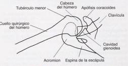
Visible Anatomical Structures
Should clearly observe:
- Lateral view of proximal humerus in relation to scapulohumeral cavity
- Coracoid process of scapula in profile
- Lesser tubercle of humerus in profile
- Scapular spine at border below joint
- Humeral head-glenoid cavity relationship
Plate Size and Orientation
Transverse orientation for optimal proximal humerus visualization
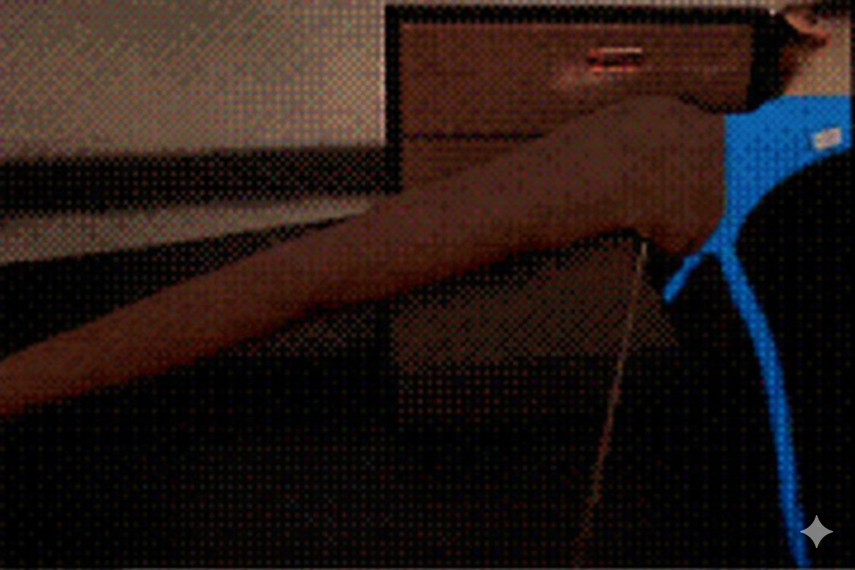
Patient Positioning
Central Ray and Angle
Direction: Directed medially 25 to 30°
Centering: Horizontally to axilla and humeral head
Adjustment: If abduction < 90°, decrease angle to 15-20°
Patient Instructions
"Hold breath and remain still"
Maintain position without movement during radiographic exposure
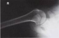
Optimal Image Characteristics
Lateral view
Proximal humerus and joint relationship
No overlap
Anatomical structures clearly differentiated
Correct profile
Coracoid process and lesser tubercle visible
Complete field
Complete scapulohumeral joint
Common Technical Challenges
Frequent problems in shoulder axial projection:
- Insufficient arm abduction (< 90°)
- Poor cassette placement away from neck
- Lack of support for arm in abduction
- Incorrect angle of central ray according to abduction degree
- Patient movement during exposure
Solution: Ensure 90° abduction when possible and adjust RC angle according to actual abduction